
Asthma‑COPD Overlap (ACO) is a clinical entity that sits between chronic obstructive pulmonary disease (COPD) and asthma, sharing features of both airway obstruction and inflammation. Recognising ACO matters because patients often experience more frequent exacerbations, a higher symptom burden, and a need for a blended therapeutic approach.
Why the Link Between COPD and Asthma Matters
Both COPD and asthma impact airflow, but they arise from different pathways. COPD, a disease most common in adults over 40, is driven largely by smokingthe inhalation of toxic particles that cause irreversible airway narrowing. Asthma, on the other hand, often begins in childhood and is linked to allergen exposuresuch as pollen, dust mites, or pet dander, triggering reversible bronchoconstriction. When a patient shows signs of both-persistent airflow limitation plus a strong eosinophilic response-they may fall into the ACO category.
Pathophysiology: Shared and Divergent Mechanisms
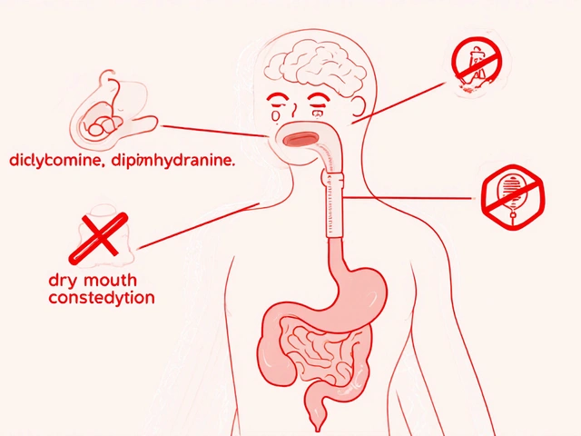
Inflammation is the common thread. In COPD, neutrophilic inflammationdominates, producing mucus hypersecretion and airway remodeling. Asthma is characterised by eosinophilic inflammationthat leads to airway hyper‑responsiveness. ACO patients often display a mixed pattern: elevated eosinophils alongside neutrophils, making diagnosis trickier but also opening doors to targeted therapies such as inhaled corticosteroids combined with long‑acting bronchodilators.
Clinical Presentation: Spotting the Overlap
- Chronic cough and sputum production (typical of COPD).
- Wheezing and episodic dyspnoea that improve with a bronchodilator (classic asthma trait).
- History of smoking plus a personal or familial atopy.
- Frequent exacerbations requiring oral steroids or hospitalisation.
Because symptoms blur, clinicians rely on objective testing to separate pure COPD, pure asthma, and ACO.
Diagnostic Toolbox: Spirometry, Biomarkers, and Imaging
Spirometrymeasures forced expiratory volume in one second (FEV1) and forced vital capacity (FVC) remains the gold standard. A post‑bronchodilator FEV1/FVC ratio below 0.70 confirms airflow obstruction. In asthma, this ratio often improves by >12% after bronchodilator; in COPD, the change is modest. ACO patients show a partial reversal-enough to hint at an asthmatic component.
Blood eosinophil countabove 300 cells/µL suggests steroid‑responsive inflammation and helps guide inhaled corticosteroid (ICS) use. High neutrophil countor C‑reactive protein (CRP) points to COPD‑dominant disease. Chest CT can reveal emphysematous changes typical of COPD, while airway wall thickening aligns with asthma.
Management Strategies: Tailoring Therapy for Overlap
Effective treatment blends the pillars of both diseases:
- Long‑acting bronchodilatorsincluding LABA (long‑acting beta‑agonists) and LAMA (long‑acting muscarinic antagonists) to keep airways open.
- Inhaled corticosteroids (ICS)reduce eosinophilic inflammation and lower exacerbation risk, especially when eosinophil counts are high.
- Short‑acting bronchodilators for rescue use during acute symptoms.
- Smoking cessation programmes, given that ongoing tobacco exposure accelerates COPD progression.
- Pulmonary rehabilitation to improve exercise tolerance and quality of life.
For patients who continue to exacerbate, biologic agents targeting interleukin‑5 (IL‑5) or IgE can be considered, mirroring asthma‑specific therapies.

Comparison: COPD vs Asthma vs ACO
| Feature | COPD | Asthma | ACO |
|---|---|---|---|
| Typical onset age | ≥40 years | Childhood-early adulthood | Mid‑life, often with prior asthma |
| Primary cause | Smoking / occupational pollutants | Allergen exposure, atopy | Combined smoking and atopy |
| Inflammation type | Neutrophilic | Eosinophilic | Mixed neutrophilic/eosinophilic |
| FEV1/FVC reversibility | ≤12% post‑bronchodilator | >12% post‑bronchodilator | Partial (5‑15%) |
| Core treatment | LAMA ± LABA | ICS+LABA | LAMA+LABA+ICS (if eosinophils high) |
Prognosis and Comorbidities
Patients with ACO tend to have a higher rate of hospitalisation compared with those having pure COPD or asthma. Cardiovascular disease, osteoporosis, and anxiety/depression are common companions, driven by chronic systemic inflammation and frequent steroid bursts. Early detection of the overlap phenotype enables clinicians to introduce ICS sooner, which can curb exacerbation frequency and potentially slow lung function decline.
Related Concepts and Next Steps
Understanding ACO opens doors to several adjunct topics that deserve a deeper dive:
- Pulmonary rehabilitationstructured exercise and education programs that improve dyspnoea and health‑related quality of life.
- Smoking cessation interventions, including nicotine replacement therapy and behavioural counselling.
- Biologic therapiestargeted drugs like mepolizumab that address eosinophilic inflammation for high‑eosinophil ACO patients.
- Use of exacerbation action planspersonalised written guides that prompt early use of oral steroids and antibiotics to prevent hospital visits.
Readers interested in the broader landscape might explore topics such as "Management of Chronic Respiratory Disease" (a broader cluster) or "Differentiating COPD Phenotypes" (a narrower, more specific article).
Bottom Line
Both COPD and asthma cause airflow trouble, but when they intertwine the result is a distinct, often more severe picture. Recognising the Asthma‑COPD overlap enables a more nuanced treatment plan, better symptom control, and a chance to halt rapid lung function loss.
Frequently Asked Questions
What is Asthma‑COPD Overlap (ACO)?
ACO is a clinical syndrome where a patient exhibits features of both chronic obstructive pulmonary disease and asthma, such as persistent airflow limitation with a partial bronchodilator response and mixed inflammatory patterns.
How can doctors tell if a patient has ACO?
Diagnosis combines spirometry (showing obstructive defect with some reversibility), blood eosinophil counts, smoking history, and symptom patterns. Imaging and response to inhaled corticosteroids also help differentiate ACO from pure COPD or asthma.
Do patients with ACO need different medication?
Yes. The typical regimen includes a long‑acting bronchodilator (LABA or LAMA) plus an inhaled corticosteroid if eosinophil levels are high. Some may benefit from biologic agents that target eosinophilic inflammation.
Is smoking still a risk factor for ACO?
Absolutely. Smoking accelerates COPD‑type changes and can worsen the overlap picture. Quitting smoking is a cornerstone of management for any ACO patient.
What is the long‑term outlook for someone with ACO?
Prognosis depends on how well the disease is controlled. Regular use of appropriate inhalers, pulmonary rehab, and avoiding triggers can reduce exacerbations and improve survival compared with untreated or misdiagnosed cases.


Emma Rauschkolb
September 25, 2025 AT 16:05Wow, the mechanistic interplay between eosinophilic‑driven bronchial hyper‑reactivity and neutrophil‑mediated airway remodeling is truly fascinating 🤓. The ACO phenotype essentially epitomizes a hybrid immunopathology, blending Th2 cytokine cascades with innate neutrophilic infiltration. Clinicians should therefore calibrate their therapeutic algorithms to target both IL‑5 pathways and mucolytic strategies. Ultimately, precision‑medicine biomarkers will dictate outcomes in this convoluted landscape.
Mara Mara
October 3, 2025 AT 02:45Indeed, the delineation of ACO demands meticulous clinical acumen, rigorous spirometric evaluation, and a steadfast commitment to evidence‑based practice!!! One cannot underestimate the pivotal role of smoking cessation, which remains the cornerstone of pulmonary preservation. Moreover, integrating long‑acting bronchodilators with inhaled corticosteroids must be executed with surgical precision. Let us champion these protocols for the betterment of our healthcare system.
Jennifer Ferrara
October 10, 2025 AT 13:25While the preceding exposition admirably underscores therapeutic rigor, an occasional orthographic imprecision may obfuscate meaning; hence, let us attend to the nuance of “inhaled corticosteroids” amid the broader pharmacologic paradigm. Moreover, the ethical imperative to tailor regimens to individual eosinophil thresholds cannot be overstated.
Terry Moreland
October 18, 2025 AT 00:05Hey folks, I totally get how confusing ACO can feel – you’re juggling asthma tricks and COPD rules at once. The key is to keep a steady inhaler routine and not skip the rehab sessions. If you notice more wheeze after a smoke break, that’s a sign to reach out to your doc fast. Stay chill and keep breathing easy!
Abdul Adeeb
October 25, 2025 AT 10:45While your encouragement is commendable, it is essential to articulate the clinical recommendations with precision: patients should undergo serial spirometric assessments, adhere to a prescribed long‑acting muscarinic antagonist, and avoid tobacco exposure unequivocally. Such rigor ensures optimal therapeutic outcomes and minimizes iatrogenic complications.
Abhishek Vernekar
November 1, 2025 AT 20:25Reading through the data, I can’t help but feel a pang of urgency 😢 – ACO patients are often caught in a therapeutic limbo, oscillating between steroids and bronchodilators. The mixed inflammatory milieu mandates both anti‑eosinophilic agents and mucolytics, lest we exacerbate systemic side effects. Let’s rally for integrated care pathways that honor both pathophysiologic arms.
Val Vaden
November 9, 2025 AT 07:05In short, ACO needs a hybrid plan – simple as that 😏.
lalitha vadlamani
November 16, 2025 AT 17:45It is profoundly disconcerting that mainstream pulmonology continues to marginalize the ACO cohort, perpetuating a narrative that trivializes their compounded morbidity. One must question the ethical complacency that allows such oversight to persist. The discourse should instead exalt vigilance, rigorous diagnostic stratification, and equitable access to biologic therapies.
kirk lapan
November 24, 2025 AT 04:25Umm… honestly, the whole “marginalization” spiel is overblown – the guidelines already address overlap, albeit succinctly. Your dramatization, while entertaining, distracts from the fact that cost‑effectiveness drives therapeutic choices. Let’s keep the conversation rooted in data, not melodrama.
Landmark Apostolic Church
December 1, 2025 AT 15:05From a sociocultural lens, ACO epitomizes the convergence of environmental exposure and genetic predisposition, a true testament to the body’s adaptive resilience. Yet the aggressiveness of smoking compounds this resilience, compelling us to adopt a more assertive public health stance.
Matthew Moss
December 9, 2025 AT 01:45While your sociocultural framing is noteworthy, it neglects the stark reality that national policies often lag in curbing tobacco proliferation, thereby imperiling citizens' pulmonary health. The moral imperative is clear: legislate stricter smoking bans and fund accessible inhaler programs. Only then can we honor the collective wellbeing of our nation.
Antonio Estrada
December 16, 2025 AT 12:25Collaboration across specialties-pulmonology, immunology, and rehabilitation-can streamline ACO management, fostering shared decision‑making and patient‑centered outcomes. By integrating objective biomarker data with personalized inhaler techniques, we reinforce therapeutic adherence and reduce exacerbation frequency.
Kevin Huckaby
December 23, 2025 AT 23:05Yo, let’s be real – the “collaboration” hype sounds nice, but bureaucratic red tape often throttles real progress 😤. If we truly want to crush ACO rates, we need bold, unapologetic funding for cutting‑edge biologics and no‑nonsense anti‑smoking crusades. 💥🚀
Brandon McInnis
December 31, 2025 AT 09:45Friends, while we debate policies and protocols, let’s not forget the human stories behind ACO – the veteran struggling with chronic cough, the teenager battling wheeze after a wildfire. These narratives remind us that every prescription, every rehab session, carries profound emotional weight. We must honor both science and soul.
Aaron Miller
January 7, 2026 AT 20:25Indeed, the sentimental veneer you employ obfuscates the stark statistics: ACO prevalence is rising at an alarming rate, and half‑hearted initiatives will not suffice!!! It is incumbent upon our nation’s leadership to deploy decisive, data‑driven interventions, not sentimental platitudes.
Roshin Ramakrishnan
January 15, 2026 AT 07:05While I appreciate the urgency conveyed, let us also cultivate inclusive dialogues that engage patients, caregivers, and policymakers alike; only through such holistic discourse can sustainable solutions emerge!!! Moreover, community‑based education programs, punctuated by culturally resonant messaging, will amplify adherence and empower individuals.
Todd Peeples
January 22, 2026 AT 17:45In the contemporary pulmonological discourse, the Asthma‑COPD Overlap (ACO) occupies a pivotal niche, demanding an integrative diagnostic algorithm that transcends the binary classification of obstructive airway diseases. Firstly, spirometric indices such as post‑bronchodilator FEV₁/FVC ratios should be interpreted in conjunction with reversibility thresholds to delineate partial bronchodilator responsiveness. Secondly, the quantification of peripheral blood eosinophil counts provides a surrogate marker for Th2‑mediated inflammation, thereby informing the judicious addition of inhaled corticosteroids to the therapeutic regimen. Thirdly, imaging modalities, particularly high‑resolution computed tomography, elucidate structural phenotypes, distinguishing emphysematous destruction from airway wall thickening. Fourthly, the incorporation of validated symptom questionnaires, for instance the COPD Assessment Test (CAT) and Asthma Control Test (ACT), facilitates longitudinal monitoring of disease burden. Fifthly, emerging biomarker panels, including periostin and fractional exhaled nitric oxide (FeNO), may further refine phenotypic stratification, although their routine clinical utility remains under investigation. Sixthly, pharmacologic stewardship mandates the synergistic deployment of long‑acting muscarinic antagonists (LAMA) and long‑acting β₂‑agonists (LABA) to ameliorate baseline airflow limitation, while inhaled corticosteroids (ICS) are reserved for those exhibiting eosinophilic predominance. Seventhly, the advent of biologic agents targeting interleukin‑5 (IL‑5) or immunoglobulin E (IgE) heralds a paradigm shift for refractory ACO cases, particularly in the context of frequent exacerbations. Eighthly, non‑pharmacologic interventions, such as pulmonary rehabilitation, confer incremental improvements in exercise tolerance and health‑related quality of life, underscoring the necessity of a multimodal approach. Ninthly, meticulous smoking cessation counseling, leveraging nicotine replacement therapies and behavioral support, remains an indispensable component of disease modification. Tenthly, regular follow‑up intervals, ideally every three to six months, enable dynamic adjustment of therapeutic intensity based on exacerbation frequency and spirometric trends. Eleventhly, vaccination strategies, encompassing annual influenza and pneumococcal immunization, mitigate infectious precipitates of acute decompensation. Twelfthly, comorbidity management, including cardiovascular risk reduction and osteoporosis screening, addresses the systemic sequelae of chronic steroid exposure. Thirteenthly, patient education initiatives, delivered through culturally tailored platforms, empower self‑management and adherence. Fourteenthly, health‑system integration, fostering seamless referrals between primary care, pulmonology, and allied health services, optimizes care continuity. Fifteenthly, future research endeavors should prioritize randomized controlled trials evaluating the comparative effectiveness of combined pharmacologic regimens in heterogeneous ACO cohorts. In summation, the confluence of rigorous phenotypic assessment, targeted pharmacotherapy, and comprehensive supportive measures constitutes the cornerstone of efficacious ACO management 😊.
Chris Smith
January 30, 2026 AT 04:25so you just listed every guideline ever written – impressive but utterly useless if nobody reads them.
Leonard Greenhall
February 6, 2026 AT 15:05While the extensive enumeration is thorough, condensing the key actionable items into a concise algorithm would enhance clinical uptake and reduce cognitive overload.