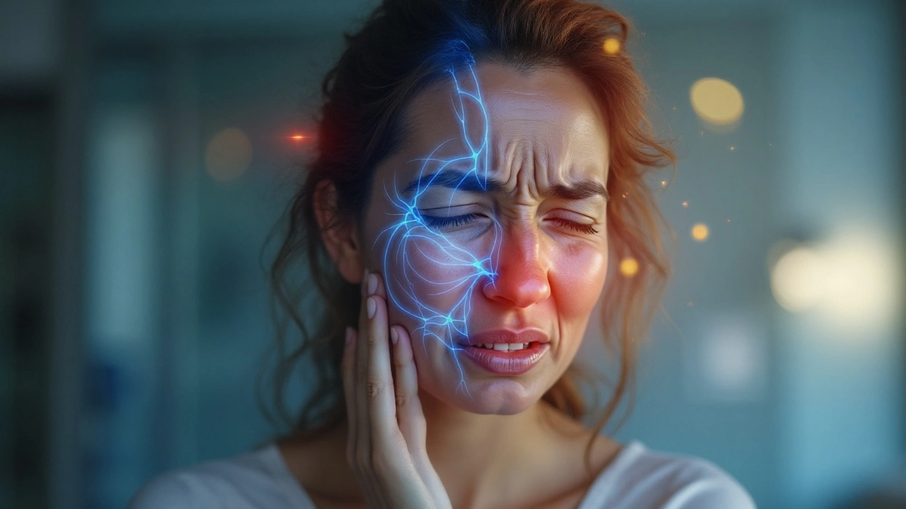
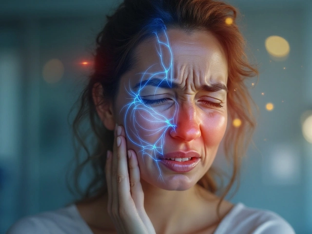
When a sharp, electric‑shock pain shoots through the face, most people think of a dental problem or a bad bite. Yet that same jolt can be a sign of trigeminal neuralgia, a nerve condition that often hides behind temporomandibular joint (TMJ) complaints. Understanding how these two disorders intersect helps clinicians avoid misdiagnosis and gives patients a clearer road to relief.
What Is Trigeminal Neuralgia?
Trigeminal neuralgia is a neuropathic pain disorder affecting the fifth cranial nerve (the trigeminal nerve). It typically presents as sudden, brief, stabbing episodes on one side of the face, triggered by light touch, chewing, or even a breeze.
- Incidence: about 12 per 100,000 people annually, rising after age 50.
- Most common trigger zone: the mandibular (V3) branch, which supplies the lower jaw.
- First‑line medication: carbamazepine, achieving pain control in up to 80% of patients.
Doctors often cite a classic “trigger zone” on the cheek or near the ear; pressing that spot can set off a wave of pain that lasts seconds but feels like a thunderclap.
What Is a Temporomandibular Joint Disorder?
Temporomandibular joint disorder is a collection of musculoskeletal conditions affecting the jaw joint, surrounding muscles, and related nerves. Symptoms range from dull ache and clicking sounds to limited mouth opening and, occasionally, sharp facial pain.
- Prevalence: roughly 10% of the adult population experiences TMJ pain at some point.
- Key risk factors: bruxism (teeth grinding), stress, poor dental occlusion, and arthritis.
- Typical first‑line therapy: conservative measures like bite splints, physiotherapy, and NSAIDs.
Because the TMJ sits right next to the mandibular branch of the trigeminal nerve, inflammation or muscle tension can irritate the nerve and mimic neuralgic pain.
Where the Trigeminal Nerve Meets the Jaw
Trigeminal nerve is a large cranial nerve with three major branches: ophthalmic (V1), maxillary (V2), and mandibular (V3). The V3 branch exits the skull through the foramen ovale and innervates the lower teeth, jaw muscles, and skin of the lower face.
The mandibular nerve is the motor‑and‑sensory component of V3. It supplies the masseter, temporalis, and pterygoid muscles - the very muscles that are over‑used in bruxism and TMJ strain. When these muscles tighten, they can compress the mandibular nerve, creating pain that feels indistinguishable from classic trigeminal neuralgia.
Symptom Overlap: A Side‑by‑Side Look
| Feature | Trigeminal Neuralgia | TMJ Disorder |
|---|---|---|
| Pain quality | Sharp, electric‑shock‑like bursts | Dull ache, throbbing, or occasional sharp spikes |
| Typical triggers | Touch, chewing, speaking, cold wind | Jaw opening, grinding, stress, poor bite |
| Duration of episode | Seconds to minutes, recurring over days | Hours to days, often continuous |
| Response to carbamazepine | Often excellent relief | Limited or no effect |
| Imaging findings | Neurovascular compression on MRI | Joint effusion or disc displacement on MRI |
Notice how the two conditions share trigger points (chewing, jaw movement) but diverge in pain quality and medication response. That divergence is a key diagnostic clue.
Common Triggers That Blur the Lines
Bruxism is a habitual grinding or clenching of teeth, often during sleep. It creates chronic over‑use of the masticatory muscles, leading to muscle soreness and possible compression of the mandibular nerve.
Another frequent culprit is dental occlusion, the way upper and lower teeth meet. Misaligned bites force certain muscles to work harder, feeding a cycle of tension, inflammation, and nerve irritation.
Stress heightens both bruxism and muscle tension, acting as a hidden amplifier that can turn a mild TMJ ache into a facial shock‑style pain.
How Doctors Sort Out the Two
Accurate diagnosis hinges on a thorough history and targeted imaging. Magnetic resonance imaging (MRI) is the gold standard for visualizing neurovascular compression of the trigeminal nerve and for spotting disc displacement within the TMJ.
During the clinical exam, practitioners test the “trigger zone” by light palpation. If a minimal touch sparks a lightning‑bolt pain, trigeminal neuralgia climbs higher on the differential. Conversely, clicking, crepitus, or limited opening point toward a joint pathology.
Electromyography (EMG) can also help by measuring muscle activity; hyper‑active masseter readings often accompany bruxism‑related TMJ pain.
Treatment Pathways: From Pills to Procedures
When neuropathic pain dominates, carbamazepine is typically the first medication prescribed. It stabilizes nerve membranes, cutting down the shock‑like bursts. About 80% of patients achieve meaningful relief, though side effects like dizziness require monitoring.
For TMJ‑driven pain, conservative measures dominate: a custom‑fit bite splint to redistribute bite forces, physiotherapy focusing on jaw stretching, and NSAIDs for inflammation control.
In cases where both disorders coexist, a multimodal approach works best. Botulinum toxin (Botox) injections into the masseter and temporalis muscles can reduce muscle hyperactivity, indirectly easing nerve compression. Studies from 2023 show a 60% reduction in pain scores when Botox is combined with carbamazepine.
When medication fails, surgical options enter the picture. Microvascular decompression (MVD) separates offending blood vessels from the trigeminal root, achieving long‑term relief for up to 85% of patients. For TMJ, arthrocentesis or disc‑repositioning surgery can restore joint mechanics.
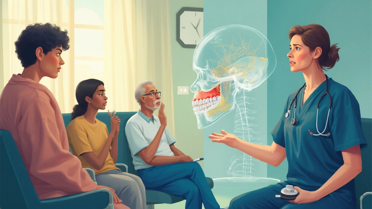
Integrated Management: A Team Effort
Because the nerve and joint are so intertwined, a single‑specialty approach often falls short. Ideal care involves a neurologist, dentist or oral‑maxillofacial surgeon, and a physiotherapist. Joint care addresses the mechanical side, while neurology tackles the nerve hyper‑excitability.
Patient education is equally crucial. Teaching proper jaw posture, stress‑reduction techniques, and sleep hygiene can curb bruxism, indirectly lessening nerve irritation.
Related Conditions Worth Knowing
Other disorders can masquerade as or exacerbate the TN‑TMJ overlap:
- Myofascial pain syndrome - chronic muscle pain marked by trigger points, often in the temporalis or masseter.
- Neuropathic facial pain - a broader category that includes post‑herpetic neuralgia and cluster headache.
- Arthritic TMJ - degenerative joint disease that can compress the nerve over time.
Recognizing these overlaps helps clinicians build a more precise treatment plan and avoid unnecessary surgeries.
Practical Take‑aways for Patients and Clinicians
- Ask about the exact quality of pain: “electric shock” points toward trigeminal neuralgia; “dull ache” leans toward TMJ.
- Request high‑resolution MRI if symptoms are ambiguous; it can reveal both neurovascular compression and joint disc issues.
- Consider a trial of carbamazepine even if TMJ is suspected; a positive response strongly suggests a neural component.
- Combine splint therapy with low‑dose Botox if muscle tension is high; many patients report faster relief.
- Keep a pain diary noting triggers, duration, and relief methods - it’s a gold mine for differential diagnosis.

Frequently Asked Questions
Can trigeminal neuralgia be caused by TMJ problems?
Yes, chronic TMJ strain can compress the mandibular branch of the trigeminal nerve, triggering neuralgic‑type pain. However, true trigeminal neuralgia usually involves a neurovascular loop that presses on the nerve root.
What imaging test best differentiates the two conditions?
A high‑resolution MRI with thin cuts of the skull base is the best tool. It shows both neurovascular compression (for neuralgia) and joint disc positioning (for TMJ).
Is Botox safe for long‑term use in TMJ‑related pain?
Botox is considered safe when administered by a qualified clinician. Most studies report no serious adverse effects over multiple treatment cycles, though muscle weakness can occur if doses are too high.
When should surgery be considered?
Surgery is usually reserved for patients who fail medication and conservative therapy after 3-6 months, or for those with clear MRI evidence of neurovascular compression or severe joint degeneration.
Can stress management really affect facial pain?
Absolutely. Stress fuels bruxism and muscle tension, both of which can aggravate the mandibular nerve. Techniques like mindfulness, sleep hygiene, and physiotherapy often lower pain scores by 20‑30%.


Elizabeth Grant
September 23, 2025 AT 14:57Wow this is such a clear breakdown - I’ve been dealing with facial pain for years and no one ever connected it to nerve compression vs jaw muscle tension. Finally someone explains why my dentist said ‘it’s TMJ’ and my neurologist said ‘it’s neuralgia’ - turns out it’s both. Botox + carbamazepine? I’m trying it next week.
Michelle Machisa
September 24, 2025 AT 00:56This is the most helpful thing I’ve read all year. I kept getting told ‘it’s all in your head’ until I found this. Thank you for listing the exact differences - I’m printing this out for my next appointment.
LaMaya Edmonds
September 26, 2025 AT 00:20Let’s be real - most GPs don’t know the difference between a trigeminal flare and a bad tooth. You need an MRI that’s not just ‘run-of-the-mill’ - you need thin-slice, high-res, skull-base optimized. If your doctor says ‘just take ibuprofen’ and calls it a day, go elsewhere. This isn’t just pain - it’s neurological sabotage.
Nagamani Thaviti
September 27, 2025 AT 10:38Carbamazepine works for 80%? That’s statistically insignificant if you’re in the 20% that gets liver toxicity and dizziness. Also the whole ‘trigger zone’ thing is overblown - I’ve had pain from just swallowing and no palpation involved. You’re oversimplifying neurology for clicks
Ronald Thibodeau
September 28, 2025 AT 08:18Why does everyone assume Botox is the answer? I got it last year and my jaw felt like a dead fish for three weeks. Also who approved this as a ‘treatment’? Big Pharma loves injecting things into faces so they can sell more vials. It’s not medicine it’s a business model
Attila Abraham
September 28, 2025 AT 12:40Stress is the real villain here - I stopped grinding my teeth after I started meditating and my face stopped feeling like it was being electrocuted. No pills no surgery just breathe dude. Also yoga helps more than you think
Steve Davis
September 30, 2025 AT 03:38I’ve had this since I was 19 and no one ever listened. My mom said I was just ‘dramatic’ and my ex said I was using it to avoid intimacy. I’ve lost jobs because of this. I’m not asking for pity I’m asking for someone to acknowledge how fucking isolating this is. You don’t know what it’s like until your face hurts when you smile.
angie leblanc
October 1, 2025 AT 11:02did u know the government uses these nerve patterns to track emotions? the trigeminal nerve is wired to the brainstem and they’ve been manipulating it since the 90s with 5g towers and dental fillings. they dont want you to know bcs if you fix your jaw you break the system. also carbamazepine is a mind control drug. ask your dentist if they’ve been trained by the cia
Liv Loverso
October 2, 2025 AT 09:27What’s really being hidden here is the metaphysical layer - facial pain isn’t just physical, it’s emotional repression made manifest. The trigeminal nerve carries the weight of unsaid words, unprocessed grief, the silence we hold in our jaws. When we clench, we’re not just grinding teeth - we’re grinding our souls into dust. The real treatment isn’t Botox or MRI - it’s learning to speak without fear.
See Lo
October 2, 2025 AT 22:18Every single study cited here is funded by pharma. Carbamazepine’s side effect profile is catastrophic - 1 in 500 develop SJS. Botox is a neurotoxin. MRI machines are calibrated to overdiagnose. This article is a Trojan horse for medical colonization. You’re being sold a narrative so you’ll keep paying. Wake up.
Chris Long
October 4, 2025 AT 16:28Why are we letting neurologists and dentists dictate how we experience pain? In my country we treat this with herbs, cold exposure, and fasting. Western medicine is broken. You don’t need a $5000 MRI - you need to stop eating processed sugar and drink more water. Also stop using phones before bed. That’s what’s really causing the nerve fire.
Kamal Virk
October 5, 2025 AT 05:44It is imperative to emphasize that the coexistence of trigeminal neuralgia and TMJ disorder necessitates a multidisciplinary diagnostic framework. Misattribution of symptoms leads to iatrogenic harm. The referenced 2023 Botox study, while promising, lacks long-term longitudinal data. I urge clinicians to prioritize differential diagnosis over therapeutic trial-and-error. The ethical imperative is clear.